Image

This past fall, the Duke Institute for Brain Sciences, in partnership with the Forever Learning Institute, brought alumni to campus for a day of learning about the mysterious and awe-inspiring organ sloshing around our skulls: the brain.
The half-day program included a brain prosthetic demo and a guided anatomy tour, capped off by lunch at the Washington Duke Inn.
Jo Supernaw M.Ed., lead organizer of the event and Director of Academic Engagement and Lifelong Learning at Duke, drew inspiration for the event from the Duke Science and Technology Initiative theme on Fortifying the Body and Brain.
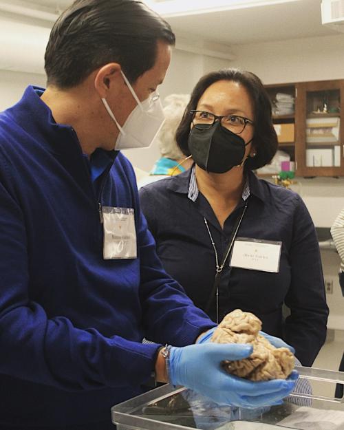
The first stop of the day brought guests to DIBS headquarters to meet with Leonard White Ph.D., the associate director of the Brain Sciences Institute, for a hands-on neuroanatomy lesson. After briefing everyone on the space, the institute, and the intentionality of the specimen donations, White led folks down the hall to the teaching lab, where they were greeted with a bevy of human brains to learn from and interact with (safety gloves required, of course). Over the course of an hour, White guided alumni through a condensed masterclass on neuroanatomy, with breaks in between for guests to examine and hold the preserved tissue.
Following the guide brain tour, participants walked a short distance to the engineering campus to learn about the cutting-edge brain technology being developed at Duke.

Jonathan Viventi Ph.D., assistant professor of biomedical engineering, and Greg Cogan, Ph.D., assistant professor of neurology, welcomed the tour group with presentations on two recent projects they’ve collaborated on to improve brain-interfacing technology.
Doctoral student Katrina Barth and graduate student Mackenna Hill walked guests through their work developing an incredibly thin and flexible activity monitor that can placed on the surface of the brain and may help to improve epilepsy diagnosis. On the other side of the room, graduate student Suseendrakumar Duraivel showed the group a snapshot of his work using electrodes to map speech areas in the brain. He played recordings of patients producing speech during their awake surgery trials, which he uses to map the course of language. These technologies are yet another stride toward a direct pathway of communication from electrical signals in the brain straight to a device.
A full morning of exploring the brain and its newest biomedical innovations left participating alumni with a clear understanding of Duke’s up-and-coming research in the neurosciences and an appreciation for the intricate organ at the center of the institute.
1 of 3
Graduate student Suseendrakumar Duraivel presents his project on electrode mapping for speech production in the brain.

1 of 3
Duke alumni get a hands-on chance to test the flexibility of doctoral student Katrina Barth’s paper-thin brain activity monitor.
1 of 3
Doctoral student Katrina Barth explains her recent research engineering a brain activity monitor placed on the surface of the brain.